実際の 6LU7 バッチジョブ(3 つの drug-like SMILES)からの公開例。ホームページで自分の構造を実行すると、ZIP と PDF エクスポート付きの非公開レポートが得られます。
自分のタンパク質をドッキング結果 ZIP には何が含まれますか?
完了した各ドッキングジョブには、ページ上のレポートと PDF に加えて results.zip が含まれます。PyMOL/Chimera、オフライン表、再現性に ZIP を使用 — PDF は同じ実行の人間可読サマリーです。
ジョブルート(実行ごとに1回)
- result.json
- ドッキング済みリガンド、失敗、スコア、redock 状態のサマリー。
- run_manifest.json
- 再現性記録:パイプラインバージョン、Vina 設定、ボックス、pH、受容体鎖、警告。
- receptor_clean.pdb
- ドッキングに使用した前処理受容体(剛性、クリーンな PDB)。
- redock_report.json
- 共結晶 redock RMSD チェック(単一リガンド / 有効時)。
ドッキング成功リガンドごと
- poses/{ligand}.sdf
- 上位3 Vina ポーズを多レコード SDF として。
- complexes/{ligand}_complex.pdb
- 3D ビューア用タンパク質–リガンド複合体。
- ligands/{ligand}/
- リガンド PDBQT、ドッキング済み PDBQT、プロトン化 SMILES。
- admet/{ligand}.json
- RDKit Lipinski、Veber、QED および関連フラグ。
- interactions/{ligand}.json
- PLIP 相互作用表(または距離ヒューリスティックフォールバック)。
- figures/{ligand}/
- 2D 相互作用図、結合部位概要 PNG、リガンド 2D 構造;PyMOL 有効時は pymol_binding_site.png オプション。
PDF レポート:親和性、ポーズ QC、印刷用に埋め込まれた相互作用図 — ZIP 内の構造ファイルの代替ではありません。
分子ドッキングレポート
サンプルドッキングレポート · PDB 6LU7 Mpro
SARS-CoV-2 メインプロテアーゼに対する3つのdrug-like SMILESのバッチスクリーニング
完了 2026/6/15 09:42:21
3
送信数
3
ドッキング成功
0
ドッキング失敗
-6.21
最良アフィニティ (kcal/mol)
トップヒット: Nirmatrelvir (-6.21 kcal/mol)
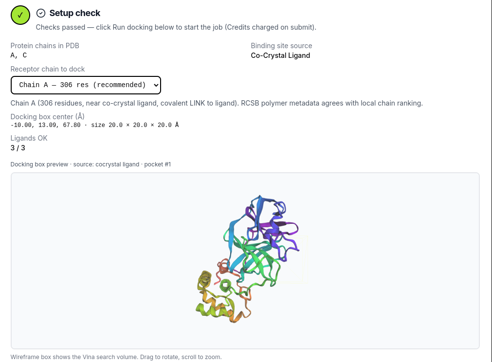
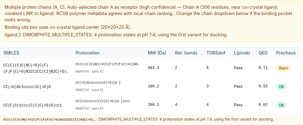
クレジット不要の検証ステップ:計算使用前にチェーン、結合部位ボックス、リガンド事前チェックを確認。
- Screening mode
- 1 受容体、3 リガンド(バッチジョブ)
- 受容体
- 6LU7 · SARS-CoV-2 メインプロテアーゼ(Mpro)
- チェーン
- A
- プロトン化 pH
- 7.4
- 結合部位
- cocrystal ligand
- サーチボックス中心 (Å)
- -10.00, 13.09, 67.80
- サーチボックスサイズ (Å)
- 20.0 × 20.0 × 20.0
再現性の詳細、ソフトウェアバージョン、査読者向けの原稿引用情報。
パイプライン (v1.9.4): Meeko 受容体/リガンド前処理と RDKit 3D 埋め込みを用いた剛性受容体 AutoDock Vina ドッキング。
プロトン化: dimorphite-DL at pH 7.4. Protonation is applied before 3D embedding and minimization (MMFF94 with UFF fallback; multi-seed ETKDG).
リガンド前処理: SMILES → dimorphite_dl → ETKDG(マルチシード)→ MMFF94/UFF → meeko リガンド準備
AutoDock Vina: exhaustiveness=8, num_modes=9, energy_range=3 kcal/mol, scoring=vina.
オープンソースソフトウェアスタック
このジョブに記録された正確なバージョン — Methods と参考文献リストで引用してください。
| コンポーネント | バージョン | 開発者 / 組織 | 役割 | Ref. |
|---|---|---|---|---|
| AutoDock Vina | 1.2.7 | Scripps Research / community fork | 剛体受容体の分子ドッキング | [2] |
| Meeko | 0.7.1 | Scripps Research (Forli lab) | 受容体・リガンドの PDBQT 前処理 | [3] |
| RDKit | 2025.3.6 | Open-source cheminformatics toolkit | SMILES → 3D 埋め込み(ETKDG)と MMFF94 最小化 | [4] |
| PLIP | 3.0.0 | University of Hamburg (Salokas et al.) | タンパク質–リガンド相互作用解析 | [5] |
| PoseBusters | 0.6.5 | University of Oxford (Buttenschoen et al.) | ドッキングポーズ品質検査(dock 設定) | [6] |
| dimorphite-DL | 2.0.2 | Open-source protonation enumerator | ジョブ pH でのリガンドプロトン化状態 | [7] |
| Biopython | 1.87 | Open-source bioinformatics library | 構造解析と受容体処理 | [8] |
| pocketeer | 0.3.1 | Apo binding-site prediction | Apo 受容体の予測ポケット順位付け | [9] |
| PyMOL (open-source) | not recorded | Schrödinger / community build | 任意の論文向け結合部位レンダリング | [10] |
論文での本ツールの引用方法
以下の段落を Methods セクションに貼り付けてください。番号付き参考文献は後に続きます。
Suggested citation paragraph (Methods / Computational details)

Molecular docking was performed using the cloud-based workflow at MolecularDocking.online (pipeline v1.9.4)[1], utilizing AutoDock Vina 1.2.7[2] with Meeko receptor/ligand preparation (Meeko 0.7.1)[3]. The receptor structure was prepared from PDB 6LU7 (chain A). The search grid was centered at (-10.00, 13.09, 67.80) Å with dimensions 20.0 × 20.0 × 20.0 Å (source: cocrystal ligand). Ligands were built from SMILES using RDKit 2025.3.6 (ETKDG embedding, MMFF94 minimization)[4] with dimorphite-DL 2.0.2 protonation at pH 7.4[7]. AutoDock Vina was run with exhaustiveness=8, num_modes=9, and energy_range=3 kcal/mol; the top-ranked pose(s) per ligand were exported. Protein–ligand interactions were analyzed with PLIP 3.0.0[5]. Pose quality was validated with PoseBusters 0.6.5 (University of Oxford docking configuration)[6]. Computational binding scores and ADMET estimates are model predictions only; experimental validation is required.

References

[1] MolecularDocking.online. Cloud molecular docking workflow (https://molecular-docking.online). Accessed 2026.
[2] Eberhardt J, Santos-Martins D, Forli S, Olson AJ. AutoDock Vina 1.2.0: new docking methods, expanded force field, and Python bindings. J Chem Inf Model. 2021;61(8):3891-3898.
[3] Forli S, Huey R, Pique ME, Sanner MF, Olson AJ. Computational protein-ligand docking and virtual drug screening with the AutoDock suite. Nat Protoc. 2016;11(5):905-919.
[4] RDKit: Open-source cheminformatics. https://www.rdkit.org/ (accessed 2024).
[5] Salokas LM, Viswanath S, Aho AJ, et al. PLIP 2021: expanding the scope of the protein-ligand interaction profiler to DNA and RNA. Nucleic Acids Res. 2023;51(W1):W530-W534.
[6] Buttenschoen GF, Morris GM, Deane CM. PoseBusters: AI-based docking methods fail to generate physically valid poses or cause unintended bias. Nat Methods. 2024;21(3):472-480.
[7] Swain M. dimorphite-DL: an open-source program for enumerating the ionization states of drug-like small molecules. J Cheminform. 2019;11(1):14.
[8] Cock PA, Antao T, Chang JT, et al. Biopython: freely available Python tools for computational molecular biology and bioinformatics. Bioinformatics. 2009;25(11):1422-1423.
[9] pocketeer: apo binding-site prediction tool used for pocket ranking in this pipeline.
[10] Schrödinger LLC. The PyMOL Molecular Graphics System, Version 2.0. (Open-source build when enabled in Advanced options.)
完全な参考文献リスト(10 件)
- MolecularDocking.online. MolecularDocking.online. Cloud molecular docking workflow (https://molecular-docking.online). Accessed 2026.
- AutoDock Vina. Eberhardt J, Santos-Martins D, Forli S, Olson AJ. AutoDock Vina 1.2.0: new docking methods, expanded force field, and Python bindings. J Chem Inf Model. 2021;61(8):3891-3898.
- Meeko / AutoDock suite. Forli S, Huey R, Pique ME, Sanner MF, Olson AJ. Computational protein-ligand docking and virtual drug screening with the AutoDock suite. Nat Protoc. 2016;11(5):905-919.
- RDKit. RDKit: Open-source cheminformatics. https://www.rdkit.org/ (accessed 2024).
- PLIP. Salokas LM, Viswanath S, Aho AJ, et al. PLIP 2021: expanding the scope of the protein-ligand interaction profiler to DNA and RNA. Nucleic Acids Res. 2023;51(W1):W530-W534.
- PoseBusters. Buttenschoen GF, Morris GM, Deane CM. PoseBusters: AI-based docking methods fail to generate physically valid poses or cause unintended bias. Nat Methods. 2024;21(3):472-480.
- dimorphite-DL. Swain M. dimorphite-DL: an open-source program for enumerating the ionization states of drug-like small molecules. J Cheminform. 2019;11(1):14.
- Biopython. Cock PA, Antao T, Chang JT, et al. Biopython: freely available Python tools for computational molecular biology and bioinformatics. Bioinformatics. 2009;25(11):1422-1423.
- pocketeer. pocketeer: apo binding-site prediction tool used for pocket ranking in this pipeline.
- PyMOL. Schrödinger LLC. The PyMOL Molecular Graphics System, Version 2.0. (Open-source build when enabled in Advanced options.)
予測結合のディスカッション形式の概要(原稿サポート段落に類似)。
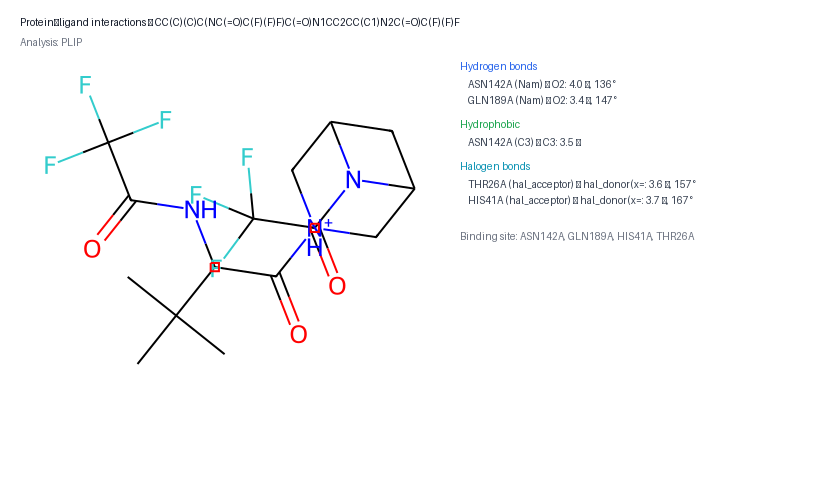
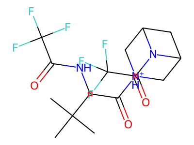
AutoDock Vinaは CC(C)(C)C(NC(=O)C(F)(F)F)C(=O)N1CC2CC(C1)N2C(=O)C(F)(F)F に対して中程度の結合を予測しました(最高スコア -6.21 kcal/mol)。 スクリーニングされた化合物の中で、CC(C)(C)C(NC(=O)C(F)(F)F)C(=O)N1CC2CC(C1)N2C(=O)C(F)(F)F がトップヒットとしてランク付けされました。 主要な予測相互作用(PLIP)には ASN142A, GLN189A との 2 個の水素結合; ASN142A との疎水性接触; HIS41A, THR26A とのハロゲン結合. リガンドは ASN142A, GLN189A, HIS41A, THR26A などのポケット残基に囲まれています。 複数のドッキングモードが類似したスコアを示しています。構造ファイル内の代替ポーズの確認を検討してください。 これらの結果は計算上の予測に過ぎず、機構的結論を導く前に実験的検証(例:SPR、ITC、細胞ベースアッセイ)が必要です。
最良モードのリードは 0.14 kcal/mol(<0.25 kcal/mol)です。順位は1位ですが、構造ファイルで近いポーズを確認してください。
PoseBusters:22/22 件のチェック合格。
PoseBusters(オックスフォード大学ドッキング設定)で検証。
検出された接触(合計 5)
PLIP- 2 個の水素結合
- 1 個の疎水接触
- 2 個のハロゲン結合
ポケット残基: ASN142A, GLN189A, HIS41A, THR26A
スライドと原稿用の PNG(ZIP にも含まれます)。
2D 相互作用図
PLIP が検出した水素結合・疎水接触・塩橋の LigPlot 風マップ。
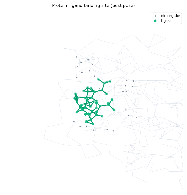
結合部位の概要
ドッキングしたリガンド付きポケット強調(matplotlib 3D 概要)。
リガンド 2D 構造
ドッキングに用いたプロトン化リガンドの化学構造。
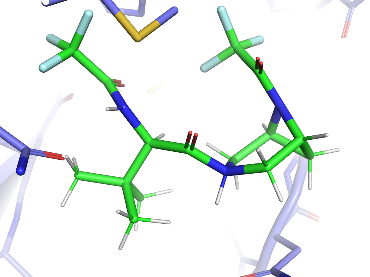
PyMOL 結合部位レンダー
レイトレースの論文用図(cartoon 受容体 + sticks)。バッチでは最良ヒットのみ。
インタラクティブなタンパク質–リガンド複合体。リガンドと Vina ポーズ(上位 3 つをエクスポート)を選択。ポーズ 1 が最高スコアのモードです。
Drag to rotate · scroll to zoom · right-drag to pan. Top 3 Vina poses are exported when available; pose 1 is the best-scoring mode.
RDKit ベースの drug-likeness 推定(Lipinski、Veber、QED)。実験 ADMET ではありません。
- 分子量 (Da)
- 404.3
- LogP
- 0.04
- TPSA (Ų)
- 70.9
- 水素結合ドナー / 水素結合アクセプター
- 2 / 3
- 回転可能結合
- 2
- QED
- 0.639
- Lipinski
- 合格
- Veber
- 合格
RDKit 記述子による計算 ADMET 推定であり、実験 ADMET データではありません。
各行はこのバッチの 1 化合物(合計 3)です。ステータス、次にアフィニティでソート。 スクリーニング比較用に ADMET 列を含みます。
| # | 化合物 | SMILES | ステータス | 最良アフィニティ (kcal/mol) | 2 位との差 | ポーズ信頼度 | Pose QC | 分子量 (Da) | Lipinski | QED |
|---|---|---|---|---|---|---|---|---|---|---|
| 1 | Nirmatrelvir | CC(C)(C)C(NC(=O)C(F)(F)F)C(=O)N1CC2CC(C1)N2C(=O)… | OK | -6.21 | 0.14 | 上位ポーズが接近 | PoseBusters QC 合格 | 404.3 | 合格 | 0.64 |
| 2 | Ibuprofen | CC(C)Cc1ccc(C(C)C(=O)O)cc1 | OK | -6.03 | 0.23 | 上位ポーズが接近 | PoseBusters QC 合格 | 205.3 | 合格 | 0.75 |
| 3 | Aspirin | CC(=O)Oc1ccccc1C(=O)O | OK | -5.05 | 0.40 | 中程度の分離 | PoseBusters QC 合格 | 179.2 | 合格 | 0.47 |
使用受容体チェーン:A
サーチボックスは共結晶リガンド中心を基準(20×20×20 Å)。
ligand_1:DIMORPHITE_MULTIPLE_STATES:pH 7.4 で 4 種類のプロトン化状態;ドッキングは最初の変異体を使用。
ligand_1:検証済みリガンド PDBQT を再利用(Review setup からプロトン化状態をロック)。
ligand_2:検証済みリガンド PDBQT を再利用(Review setup からプロトン化状態をロック)。
ligand_3:検証済みリガンド PDBQT を再利用(Review setup からプロトン化状態をロック)。
- 剛性受容体のみ(側鎖は柔軟化しない)。
- ジョブあたり 1 タンパク質チェーン(複数チェーン PDB はチェーンを選択)。
- リガンドプロトン化は dimorphite_dl をジョブ pH(デフォルト 7.4)またはリガンドごとの pH 上書き(4.0–10.0)で実行。
- リガンド入力:SMILES、MOL ブロック、または SDF(最初のレコード)。
- オプション preserve_hetatm(例 ZN、MG)はクリーンな受容体 PDB で金属を保持。
- 結合リガンド検出時に共結晶 redock sanity チェック(run_redock=true)。
Ready to run your own docking job?
Start on the home page