Ví dụ công bố từ công việc batch 6LU7 thực (ba SMILES giống thuốc). Chạy cấu trúc của bạn trên trang chủ để nhận báo cáo riêng với ZIP và PDF tải xuống.
Docking protein của bạnZIP kết quả có gì?
Mỗi job docking hoàn tất đều có results.zip cùng báo cáo trên trang và PDF. Dùng ZIP cho PyMOL/Chimera, bảng offline và tái lập — PDF là bản tóm tắt dễ đọc của cùng một lần chạy.
Gốc job (một lần mỗi run)
- result.json
- Tóm tắt ligand đã dock, thất bại, điểm số và trạng thái redock.
- run_manifest.json
- Bản ghi tái lập: phiên bản pipeline, cài đặt Vina, box, pH, chuỗi receptor, cảnh báo.
- receptor_clean.pdb
- Receptor đã chuẩn bị dùng cho docking (PDB cứng, đã làm sạch).
- redock_report.json
- Kiểm tra RMSD redock đồng tinh thể (ligand đơn / khi bật).
Mỗi ligand dock thành công
- poses/{ligand}.sdf
- Top 3 pose Vina dưới dạng SDF nhiều bản ghi.
- complexes/{ligand}_complex.pdb
- Complex protein–ligand cho trình xem 3D.
- ligands/{ligand}/
- PDBQT ligand, PDBQT đã dock và SMILES đã proton hóa.
- admet/{ligand}.json
- RDKit Lipinski, Veber, QED và các cờ liên quan.
- interactions/{ligand}.json
- Bảng tương tác PLIP (hoặc fallback heuristic khoảng cách).
- figures/{ligand}/
- Sơ đồ tương tác 2D, PNG tổng quan vùng liên kết, cấu trúc 2D ligand; pymol_binding_site.png tùy chọn nếu bật PyMOL.
Báo cáo PDF: affinity, QC pose, hình tương tác nhúng để in — không thay thế file cấu trúc trong ZIP.
Báo cáo docking phân tử
Báo cáo mẫu · PDB 6LU7 Mpro
Sàng lọc theo lô ba SMILES giống thuốc chống protease chính SARS-CoV-2
Hoàn thành 2026/6/15 09:42:21
3
Đã gửi
3
Docking thành công
0
Không thể docking
-6.21
Affinity tốt nhất (kcal/mol)
Kết quả hàng đầu: Nirmatrelvir (-6.21 kcal/mol)
Bước xác thực không tính credit: xác nhận chuỗi, hộp vị trí liên kết và precheck ligand trước khi dùng tính toán.
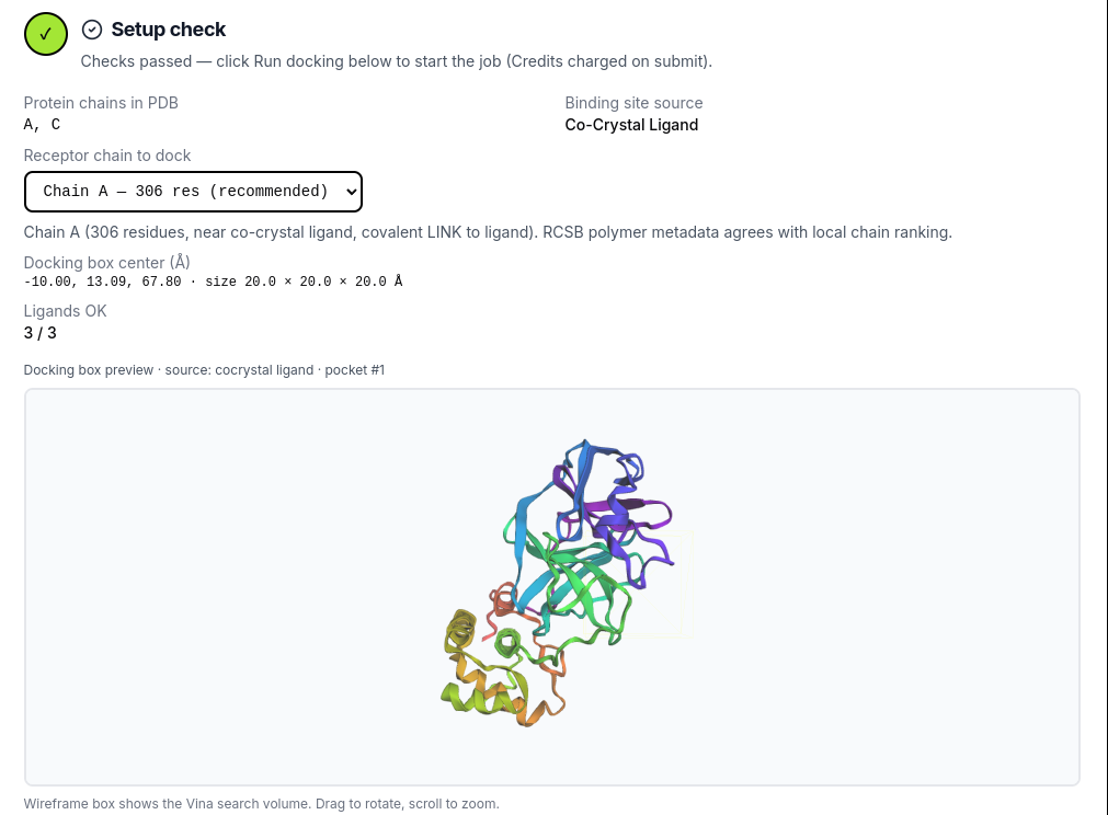
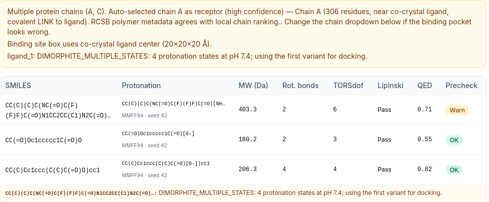
- Screening mode
- Một thụ thể, 3 ligand (công việc theo lô)
- Thụ thể
- 6LU7 · Protease chính SARS-CoV-2 (Mpro)
- Chuỗi
- A
- pH proton hóa
- 7.4
- Vị trí liên kết
- cocrystal ligand
- Tâm hộp tìm kiếm (Å)
- -10.00, 13.09, 67.80
- Kích thước hộp tìm kiếm (Å)
- 20.0 × 20.0 × 20.0
Chi tiết tái lập, phiên bản phần mềm và trích dẫn sẵn sàng cho bản thảo cho phản biện.
Pipeline (v1.9.4): Docking AutoDock Vina thụ thể cứng với chuẩn bị thụ thể/ligand Meeko và embedding 3D RDKit.
Proton hóa: dimorphite-DL at pH 7.4. Protonation is applied before 3D embedding and minimization (MMFF94 with UFF fallback; multi-seed ETKDG).
Chuẩn bị ligand: SMILES → dimorphite_dl → ETKDG (đa seed) → MMFF94/UFF → meeko_prepare_ligand
AutoDock Vina: exhaustiveness=8, num_modes=9, energy_range=3 kcal/mol, scoring=vina.
Stack phần mềm mã nguồn mở
Phiên bản chính xác được ghi cho công việc này — trích dẫn trong Methods và danh mục tài liệu.
| Thành phần | Phiên bản | Nhà phát triển / tổ chức | Vai trò | Ref. |
|---|---|---|---|---|
| AutoDock Vina | 1.2.7 | Scripps Research / community fork | Docking phân tử thụ thể cứng | [2] |
| Meeko | 0.7.1 | Scripps Research (Forli lab) | Chuẩn bị PDBQT thụ thể và ligand | [3] |
| RDKit | 2025.3.6 | Open-source cheminformatics toolkit | SMILES → nhúng 3D (ETKDG) và tối thiểu hóa MMFF94 | [4] |
| PLIP | 3.0.0 | University of Hamburg (Salokas et al.) | Phân tích tương tác protein–ligand | [5] |
| PoseBusters | 0.6.5 | University of Oxford (Buttenschoen et al.) | Kiểm tra chất lượng pose docking (cấu hình dock) | [6] |
| dimorphite-DL | 2.0.2 | Open-source protonation enumerator | Trạng thái proton hóa ligand ở pH của job | [7] |
| Biopython | 1.87 | Open-source bioinformatics library | Phân tích cấu trúc và xử lý thụ thể | [8] |
| pocketeer | 0.3.1 | Apo binding-site prediction | Xếp hạng túi dự đoán trên thụ thể apo | [9] |
| PyMOL (open-source) | not recorded | Schrödinger / community build | Render tùy chọn vị trí gắn kết cho công bố | [10] |
Cách trích dẫn công cụ này trong bài báo
Dán đoạn văn bên dưới vào phần Methods; tài liệu tham khảo đánh số theo sau.
Suggested citation paragraph (Methods / Computational details)

Molecular docking was performed using the cloud-based workflow at MolecularDocking.online (pipeline v1.9.4)[1], utilizing AutoDock Vina 1.2.7[2] with Meeko receptor/ligand preparation (Meeko 0.7.1)[3]. The receptor structure was prepared from PDB 6LU7 (chain A). The search grid was centered at (-10.00, 13.09, 67.80) Å with dimensions 20.0 × 20.0 × 20.0 Å (source: cocrystal ligand). Ligands were built from SMILES using RDKit 2025.3.6 (ETKDG embedding, MMFF94 minimization)[4] with dimorphite-DL 2.0.2 protonation at pH 7.4[7]. AutoDock Vina was run with exhaustiveness=8, num_modes=9, and energy_range=3 kcal/mol; the top-ranked pose(s) per ligand were exported. Protein–ligand interactions were analyzed with PLIP 3.0.0[5]. Pose quality was validated with PoseBusters 0.6.5 (University of Oxford docking configuration)[6]. Computational binding scores and ADMET estimates are model predictions only; experimental validation is required.

References

[1] MolecularDocking.online. Cloud molecular docking workflow (https://molecular-docking.online). Accessed 2026.
[2] Eberhardt J, Santos-Martins D, Forli S, Olson AJ. AutoDock Vina 1.2.0: new docking methods, expanded force field, and Python bindings. J Chem Inf Model. 2021;61(8):3891-3898.
[3] Forli S, Huey R, Pique ME, Sanner MF, Olson AJ. Computational protein-ligand docking and virtual drug screening with the AutoDock suite. Nat Protoc. 2016;11(5):905-919.
[4] RDKit: Open-source cheminformatics. https://www.rdkit.org/ (accessed 2024).
[5] Salokas LM, Viswanath S, Aho AJ, et al. PLIP 2021: expanding the scope of the protein-ligand interaction profiler to DNA and RNA. Nucleic Acids Res. 2023;51(W1):W530-W534.
[6] Buttenschoen GF, Morris GM, Deane CM. PoseBusters: AI-based docking methods fail to generate physically valid poses or cause unintended bias. Nat Methods. 2024;21(3):472-480.
[7] Swain M. dimorphite-DL: an open-source program for enumerating the ionization states of drug-like small molecules. J Cheminform. 2019;11(1):14.
[8] Cock PA, Antao T, Chang JT, et al. Biopython: freely available Python tools for computational molecular biology and bioinformatics. Bioinformatics. 2009;25(11):1422-1423.
[9] pocketeer: apo binding-site prediction tool used for pocket ranking in this pipeline.
[10] Schrödinger LLC. The PyMOL Molecular Graphics System, Version 2.0. (Open-source build when enabled in Advanced options.)
Danh sách tham khảo đầy đủ (10 mục)
- MolecularDocking.online. MolecularDocking.online. Cloud molecular docking workflow (https://molecular-docking.online). Accessed 2026.
- AutoDock Vina. Eberhardt J, Santos-Martins D, Forli S, Olson AJ. AutoDock Vina 1.2.0: new docking methods, expanded force field, and Python bindings. J Chem Inf Model. 2021;61(8):3891-3898.
- Meeko / AutoDock suite. Forli S, Huey R, Pique ME, Sanner MF, Olson AJ. Computational protein-ligand docking and virtual drug screening with the AutoDock suite. Nat Protoc. 2016;11(5):905-919.
- RDKit. RDKit: Open-source cheminformatics. https://www.rdkit.org/ (accessed 2024).
- PLIP. Salokas LM, Viswanath S, Aho AJ, et al. PLIP 2021: expanding the scope of the protein-ligand interaction profiler to DNA and RNA. Nucleic Acids Res. 2023;51(W1):W530-W534.
- PoseBusters. Buttenschoen GF, Morris GM, Deane CM. PoseBusters: AI-based docking methods fail to generate physically valid poses or cause unintended bias. Nat Methods. 2024;21(3):472-480.
- dimorphite-DL. Swain M. dimorphite-DL: an open-source program for enumerating the ionization states of drug-like small molecules. J Cheminform. 2019;11(1):14.
- Biopython. Cock PA, Antao T, Chang JT, et al. Biopython: freely available Python tools for computational molecular biology and bioinformatics. Bioinformatics. 2009;25(11):1422-1423.
- pocketeer. pocketeer: apo binding-site prediction tool used for pocket ranking in this pipeline.
- PyMOL. Schrödinger LLC. The PyMOL Molecular Graphics System, Version 2.0. (Open-source build when enabled in Advanced options.)
Tóm tắt kiểu thảo luận về liên kết dự đoán (tương tự hỗ trợ bản thảo).
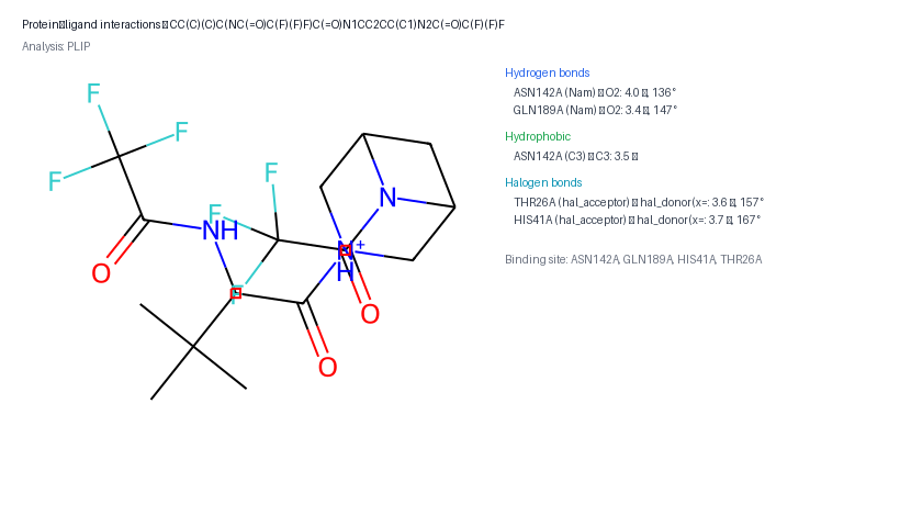
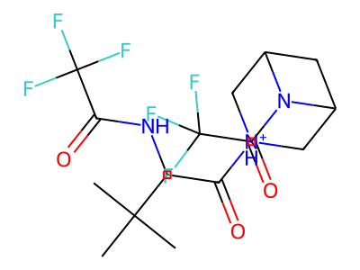
AutoDock Vina dự đoán liên kết vừa phải cho CC(C)(C)C(NC(=O)C(F)(F)F)C(=O)N1CC2CC(C1)N2C(=O)C(F)(F)F (điểm tốt nhất -6.21 kcal/mol). Trong số các hợp chất được sàng lọc, CC(C)(C)C(NC(=O)C(F)(F)F)C(=O)N1CC2CC(C1)N2C(=O)C(F)(F)F xếp hạng cao nhất. Các tương tác chính dự đoán (PLIP) bao gồm 2 liên kết hydro với ASN142A, GLN189A; tiếp xúc kỵ nước liên quan đến ASN142A; liên kết halogen với HIS41A, THR26A. Ligand được bao quanh bởi các residue trong túi, bao gồm ASN142A, GLN189A, HIS41A, THR26A. Nhiều chế độ docking có điểm tương tự; hãy xem xét kiểm tra các pose thay thế trong các tệp cấu trúc. Kết quả này chỉ là dự đoán tính toán và cần được xác minh bằng thực nghiệm (ví dụ: SPR, ITC hoặc thử nghiệm trên tế bào) trước khi rút ra kết luận cơ chế.
Mode tốt nhất dẫn trước 0.14 kcal/mol (<0.25 kcal/mol). Vẫn xếp hạng nhất nhưng nên xem lại các pose gần trong file cấu trúc.
PoseBusters: 22/22 kiểm tra đạt.
Xác thực bằng PoseBusters (cấu hình docking của Đại học Oxford).
Tiếp xúc phát hiện (5 tổng)
PLIP- 2 liên kết hydro
- 1 tiếp xúc kỵ nước
- 2 liên kết halogen
Phần tử trong pocket: ASN142A, GLN189A, HIS41A, THR26A
Ảnh PNG sẵn dùng cho slide và bản thảo (cũng có trong ZIP).
Sơ đồ tương tác 2D
Bản đồ kiểu LigPlot về liên kết hydro, tiếp xúc kỵ nước và cầu muối do PLIP phát hiện.
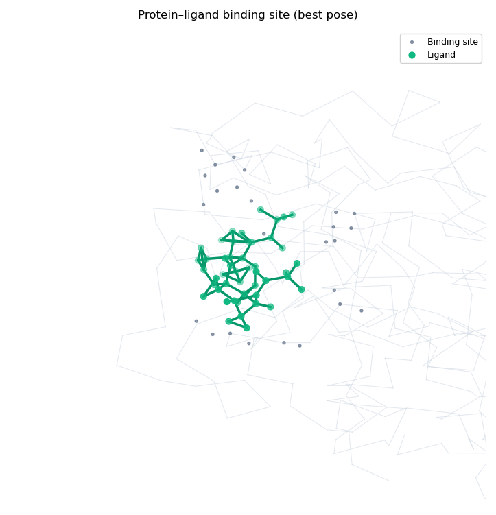
Tổng quan vị trí liên kết
Túi được làm nổi với ligand đã dock (tổng quan 3D matplotlib).
Cấu trúc ligand 2D
Cấu trúc hóa học của ligand đã proton hóa dùng cho docking.
Hình render PyMOL vị trí liên kết
Hình công bố ray-traced (thụ thể cartoon + sticks). Chỉ hit tốt nhất với batch.
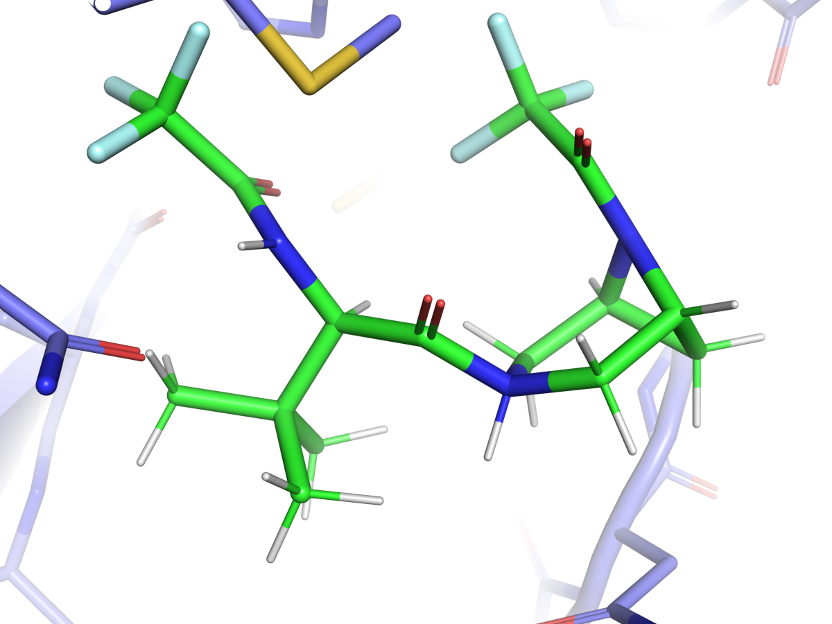
Phức hợp protein–ligand tương tác. Chọn ligand và pose Vina (3 pose hàng đầu được xuất). Pose 1 là mode có điểm cao nhất.
Drag to rotate · scroll to zoom · right-drag to pan. Top 3 Vina poses are exported when available; pose 1 is the best-scoring mode.
Ước tính drug-likeness dựa trên RDKit (Lipinski, Veber, QED). Không phải ADMET thự nghiệm.
- Khối lượng phân tử (Da)
- 404.3
- LogP
- 0.04
- TPSA (Ų)
- 70.9
- Donor liên kết H / Acceptor liên kết H
- 2 / 3
- Liên kết xoay
- 2
- QED
- 0.639
- Lipinski
- Đạt
- Veber
- Đạt
Ước tính ADMET tính toán từ mô tả RDKit, không phải dữ liệu ADMET thự nghiệm.
Mỗi hàng là một hợp chất trong lô này (3 tổng cộng). Sắp xếp theo trạng thái, rồi affinity. Cột ADMET được bao gồm để so sánh sàng lọc.
| # | Hợp chất | SMILES | Trạng thái | Affinity tốt nhất (kcal/mol) | Δ so với thứ 2 | Độ tin cậy pose | Pose QC | KL (Da) | Lipinski | QED |
|---|---|---|---|---|---|---|---|---|---|---|
| 1 | Nirmatrelvir | CC(C)(C)C(NC(=O)C(F)(F)F)C(=O)N1CC2CC(C1)N2C(=O)… | OK | -6.21 | 0.14 | Các pose đầu gần nhau | PoseBusters QC đạt | 404.3 | Đạt | 0.64 |
| 2 | Ibuprofen | CC(C)Cc1ccc(C(C)C(=O)O)cc1 | OK | -6.03 | 0.23 | Các pose đầu gần nhau | PoseBusters QC đạt | 205.3 | Đạt | 0.75 |
| 3 | Aspirin | CC(=O)Oc1ccccc1C(=O)O | OK | -5.05 | 0.40 | Tách biệt vừa phải | PoseBusters QC đạt | 179.2 | Đạt | 0.47 |
Chuỗi thụ thể đã dùng: A
Hộp tìm kiếm dùng tâm ligand co-tinh thể (20×20×20 Å).
ligand_1: DIMORPHITE_MULTIPLE_STATES: 4 trạng thái proton hóa ở pH 7.4; dùng biến thể đầu tiên cho docking.
ligand_1: Tái sử dụng PDBQT ligand đã xác thực (proton hóa khóa từ Review setup).
ligand_2: Tái sử dụng PDBQT ligand đã xác thực (proton hóa khóa từ Review setup).
ligand_3: Tái sử dụng PDBQT ligand đã xác thực (proton hóa khóa từ Review setup).
- Chỉ thụ thể cứng (không có side chain linh hoạt).
- Một chuỗi protein mỗi công việc (chọn chuỗi khi PDB có nhiều chuỗi).
- Proton hóa ligand dùng dimorphite_dl ở pH công việc (mặc định 7.4) hoặc ghi đè pH theo ligand (4.0–10.0).
- Đầu vào ligand: SMILES, khối MOL hoặc SDF (bản ghi đầu tiên).
- preserve_hetatm tùy chọn (vd. ZN, MG) giữ kim loại trong PDB thụ thể đã làm sạch.
- Kiểm tra sanity redock co-tinh thể khi phát hiện ligand gắn (run_redock=true).
Ready to run your own docking job?
Start on the home page